X-ray test
द्वारा सहकर्मी समीक्षा की गई Dr Colin Tidy, MRCGPद्वारा अंतिम अपडेट Dr Hayley Willacy, FRCGP अंतिम अद्यतन 21 अगस्त 2023
मरीज की संपादकीय दिशानिर्देशों
- डाउनलोडडाउनलोड
- साझा करें
- Language
- चर्चा
- ऑडियो संस्करण
- Google पर पसंदीदा स्रोतों में जोड़ें
X-ray tests show bones and certain other tissues.
एक नजर में
X-rays are a type of high-energy radiation used to create images inside the body.
Dense body parts like bones appear white, while air-filled parts appear black.
X-rays can help diagnose bone issues, heart conditions, lung infections, and swallowed items.
The test is painless and typically involves a short burst of X-rays passing through the body.
A radiologist will interpret the X-ray images and send a report to your doctor.
इस लेख में:
के लिए वीडियो चयन इमेजिंग
नीचे पढ़ना जारी रखें
What are X-rays?
X-rays are a type of high-energy radiation. An X-ray machine can produce short bursts of X-rays. The rays pass easily through fluids and soft tissues of the body. However, dense tissue such as bone will block some of the X-rays. (Density means how much of something there is in a certain amount of space.) The more dense the tissue, the fewer X-rays pass through. Air and water are less dense because the particles which make them are not held closely together. They let more X-rays pass through them.
What do chest X-rays show?
सामग्री पर वापस जाएंIn an X-ray picture bones show as light/white areas, whereas air shows as black or darker areas. The other lighter area in the middle of the image is the heart. The muscle which makes up the heart is also quite dense and stops X-rays passing through.
Posteroanterior chest X-ray
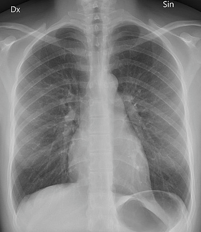
© Mikael Häggström, CC0, via Wikimedia Commons
नीचे पढ़ना जारी रखें
How is an X-ray test done?
सामग्री पर वापस जाएंA film, similar to a photographic film, is placed behind the part of the body being X-rayed. The X-ray machine fires a short burst of X-rays through part of your body. The X-rays hit the film, which is then developed. The more X-rays that hit the film, the blacker it develops. So, dense parts of the body that block many of the X-rays show up as white (such as bones). Hollow or air-filled parts of the body show up as black (such as parts of the lung). Soft tissues (such as muscle and body organs) show up as various shades of grey, depending on how dense they are.
The developed film is studied by an X-ray doctor (radiologist) who sends a report to the doctor who requested the test.
An ordinary X-ray test is painless. You cannot see or feel X-rays. You should stay still when the X-ray beam is 'fired', as otherwise the picture may be blurred.
What can an X-ray diagnose?
सामग्री पर वापस जाएंBones
Bones, teeth, bone fractures and other abnormalities of bone.
Joint spaces and some abnormalities of joints, such as osteoarthritis.
Chest
The size and shape of the heart. So, certain heart conditions can be detected.
Changes in the density of some softer tissues. For example, a lung tumour is more dense than air-filled lung and will show as a 'shadow' on a chest X-ray. A breast tumour (breast cancer) is more dense than ordinary breast tissue and shows as a 'shadow' on an X-ray of the breast. An X-ray of the breast is also known as a mammogram.
Lung infections (such as निमोनिया या tuberculosis) can be diagnosed with a chest x-ray.
Collections of fluid - for example, in the lung or gut - may show as grey 'shadows' against the normal black of the air-filled chest, or hollow gut.
Brain
Blocked blood vessels - a contrast agent that contains iodine can help make areas of your circulatory system (blood vessels) visible on X-rays. A blocked blood vessel in the brain occurs in a स्ट्रोक. This type of x-ray is called an angiogram.
Abdomen
Digestive tract problems (such as difficulty swallowing), are investigated by a barium swallow. A drink containing a contrast agent called barium, is swallowed and this shows up the outline of the gullet. Most blockages can be highlighted this way.
Accidentally swallowed items - metal, glass and stone objects show up quite well on plain x-rays. They will show the doctors where the object is and allow planning of how best to remove it. Other items (such as those made of wood, or plastic) may be seen best using other methods of imaging such as ultrasound या CT scanning.
नीचे पढ़ना जारी रखें
Types of X-rays
सामग्री पर वापस जाएंAn ordinary (often called 'plain') X-ray is a quick, easy and relatively cheap test. It may be all that is needed to diagnose or assess various problems. However, an ordinary X-ray has limited uses.
More specialised types of x-rays include:
Contrast studies: substances are used to highlight certain structures in the x-rays. Common examples are blood vessels (angiograms), different parts of the gut (barium swallows and enemas), and urinary system including the bladder and kidneys, (urograms).
Mammograms: these are special x-rays of the breasts designed to show possible cancerous areas. They are used in breast screening programmes.
CT (computerised tomography); a series of x-ray pictures from different angles which a computer puts togther to give a detailed whole picture.
DEXA (dual energy x-rays absorptiometry); a low dose of x-rays which is designed to measure bone density. This is useful when diagnosing ऑस्टियोपोरोसिस.
How do I get an X-ray?
सामग्री पर वापस जाएंYour GP or consultant may order an X-ray to help diagnose a medical problem you have been discussing with them. X-rays are usually taken in your local hospital department, although some larger health centres also now take certain simple X-rays.
A report on the X-ray is sent to the doctor who ordered the test. The report will come from a specialist in X-rays and their interpretation - a radiologist. The pictures obtained from the X-ray are not usually seen by the requesting doctor, unless it is an emergency situation such as in an A&E department.
How long do X-ray results take?
सामग्री पर वापस जाएंX-ray pictures are looked at by a specialist called a radiologist, who sends a report to the requesting doctor. This should be available to that doctor within 1-2 weeks.
If there is a serious or urgent finding the report is sent as soon as possible.
Are X-rays dangerous?
सामग्री पर वापस जाएंThere is very little risk with having one X-ray test. However, with repeated tests there is a risk that the X-rays may damage some cells in the body, possibly leading to cancer in the future. The amount of radiation is always kept to the minimum needed to obtain a good picture of the particular body part being checked. (Also, radiographers who take the X-ray pictures always wear lead aprons or go behind a protective screen when the X-rays are fired, to avoid repeated radiation exposure to X-rays.)
Can you have an X-ray while pregnant?
If possible, pregnant women should not have an X-ray test, as there is a small risk that X-rays may cause an abnormality to the unborn child. This is why women are asked if they are, or might be, pregnant, before having an X-ray.
रोगी के लिए चयन इमेजिंग

परीक्षण और जांच
पीईटी स्कैन
एक पीईटी स्कैन छवियाँ बनाता है जो दिखाती हैं कि शरीर में कहाँ कोशिकाएँ विशेष रूप से सक्रिय हैं। इसका सबसे अधिक उपयोग कैंसर का निदान और मूल्यांकन करने के लिए किया जाता है। नोट: नीचे दी गई जानकारी केवल एक सामान्य मार्गदर्शिका है। व्यवस्थाएँ और परीक्षण करने के तरीके विभिन्न अस्पतालों के बीच भिन्न हो सकते हैं। हमेशा अपने डॉक्टर या स्थानीय अस्पताल द्वारा दिए गए निर्देशों का पालन करें।.
डॉ. रोजालिन एडलमैन, MRCGP द्वारा

परीक्षण और जांच
Radionuclide scan
A radionuclide scan is a way of imaging bones, organs and other parts of the body by using a small dose of a radioactive chemical. There are different types of radionuclide chemical. The one used depends on which organ or part of the body is to be scanned. Note: the information below is a general guide only. The arrangements, and the way tests are performed, may vary between different hospitals. Always follow the instructions given by your doctor or local hospital.
डॉ. राचेल हडसन, MRCGP द्वारा
अक्सर पूछे जाने वाले प्रश्न
What is the difference between an ordinary X-ray and more specialised types?
An ordinary or 'plain' X-ray is a quick, easy, and relatively cheap test for diagnosing various problems. However, it has limited uses. More specialised X-rays, such as contrast studies, mammograms, CT scans, and DEXA scans, use additional techniques or technology to provide more detailed or specific information depending on what is being investigated.
What happens if I move during an X-ray?
It's important to stay very still when the X-ray beam is 'fired'. If you move, the picture may be blurred, which could make it difficult for the radiologist to interpret clearly.
How quickly will my GP receive the results of my X-ray?
The X-ray pictures are reviewed by a radiologist who then sends a report to the doctor who requested the test. This report should typically be available to your doctor within 1-2 weeks. If there is a serious or urgent finding, the report will be sent as soon as possible.
Where are X-rays usually performed?
X-rays are generally taken in a hospital department. Some larger health centres also have the facilities to take certain simple X-rays.
Are X-rays able to show swallowed objects?
Yes, some accidentally swallowed items can be quite visible on plain X-rays. Metal, glass, and stone objects tend to show up well, helping doctors determine their location and plan for removal. However, objects made of wood or plastic might be better seen using other imaging methods like ultrasound or CT scanning.
What is the role of a radiologist?
A radiologist is a specialist in X-rays and their interpretation. They study the developed X-ray film and send a report to the doctor who requested the test, detailing their findings.
अधिक पठन और संदर्भ
- Tafti D, Maani CV; X-ray Production.
- Frane N, Bitterman A; Radiation Safety and Protection.
नीचे पढ़ना जारी रखें
लेखक के बारे मेंपूरा जीवन परिचय देखें

Dr Hayley Willacy, FRCGP
सामान्य चिकित्सक, चिकित्सा लेखक
MBChB (1992), DRCOG, DFFP, MRCOG (Part 1) MRCGP (2007), DFSRH (2013), MSc - medical education (2020)
डॉ हेले विलासी एक एनएचएस जीपी थीं जो उत्तर पश्चिम इंग्लैंड में काम कर रही थीं, जिन्होंने 30 वर्षों के बाद 2022 में नैदानिक अभ्यास से सेवानिवृत्ति ली।.
समीक्षक के बारे मेंपूरा जीवन परिचय देखें

Dr Colin Tidy, MRCGP
सामान्य चिकित्सक, चिकित्सा लेखक
MBBS, MRCGP, MRCP (Paediatrics), DCH
डॉ. कॉलिन टिडी एक एनएचएस डॉक्टर हैं, जो ऑक्सफोर्डशायर में स्थित हैं।.
लेख का इतिहास
इस पृष्ठ पर दी गई जानकारी योग्य चिकित्सकों द्वारा लिखी और सहकर्मी समीक्षा की गई है।.
अगली समीक्षा की तिथि: 19 अगस्त 2028
21 अगस्त 2023 | नवीनतम संस्करण

पूछें, साझा करें, जुड़ें।.
चर्चाओं को ब्राउज़ करें, प्रश्न पूछें, और सैकड़ों स्वास्थ्य विषयों पर अनुभव साझा करें।.

अस्वस्थ महसूस कर रहे हैं?
अपने लक्षणों का ऑनलाइन मुफ्त में मूल्यांकन करें
पेशेंट न्यूज़लेटर के लिए साइन अप करें
आपकी साप्ताहिक खुराक स्पष्ट, विश्वसनीय स्वास्थ्य सलाह की - जो आपको सूचित, आत्मविश्वासी और नियंत्रण में महसूस करने में मदद करने के लिए लिखी गई है।.
सदस्यता लेने पर आप हमारी स्वीकार करते हैं गोपनीयता नीति. आप किसी भी समय सदस्यता समाप्त कर सकते हैं। हम कभी भी आपका डेटा नहीं बेचते।.